Lead researcher
An TangKeywods
Magnetic resonance imaging (MRI), elastography, steatosis, fibrosis, liver, abdomen, cancer, artificial intelligence.
Research interests
A. Quantitative ultrasound techniques for diagnosis of nonalcoholic steatohepatitis (NASH).
An Tang is the PI of a cross-sectional clinical imaging trial performed in collaboration at CHUM and MUHC, entitled, ‘Quantitative ultrasound (QUS) techniques for diagnosis of NASH‘ in volunteers and patients with a spectrum of NAFLD and NASH, using histopathology as the reference standard in patients. Our advanced QUS radiofrequency-based methods will include: spectral analysis, speckle properties, and viscoelastic properties. We will assess the diagnostic accuracy of QUS techniques for steatosis quantification, NASH diagnosis, and compare the diagnostic accuracy of QUS with magnetic resonance imaging (MRI) (Figure 1). Considering the non-invasiveness, safety profile, cost-effectiveness and bedside portability of ultrasound, we envision that the clinical validation of QUS-based methods will complement the capabilities of advanced MRI by providing clinicians with new point-of-care imaging techniques for better diagnosis and monitoring of therapy response in chronic liver disease.
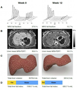
Figure 1. 57-year-old woman with type 2 diabetes before and after treatment. (A) Magnetic resonance spectroscopy (MRS), (B) liver mean MRI-proton density fat fraction (PDFF), (C) total liver volume, and (D) total liver fat index (TLFI). Tang A, et al. Diabetes care. 2015;38(7):1339-46.
B. Cancer analysis with deep learning artificial intelligence (CANDELA)
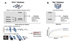
An Tang is the medical co-PI of a study entitled ‘Cancer analysis with deep Learning artificial intelligence (CANDELA)‘ involving the development and clinical application of artificial intelligence (AI) techniques applied to medical imaging. We have built an imaging database of resected liver tumors. We are evaluating the accuracy of AI methods based on deep learning algorithms for detection, segmentation (Figure 2), classification of liver tumors and prediction of clinical outcomes (Figure 3) such as overall survival and tumor-free survival. Considering that Deep Learning has recently shown human-level performance on image recognition and image labeling, we anticipate that these techniques have the potential to improve tumor detection, segmentation, diagnostic accuracy, prediction of therapeutic response and prediction of survival. The innovative technology that will be developed as part of this proposal has a high potential for transfer to clinical care and commercialization in terms of licensing of technologies.
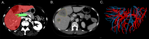
Figure 2. Prior work in (A) liver segmentation, (B) automated tumor segmentation (red) in agreement with manual segmentation by radiology experts (green), and (C) vascular segmentation of portal veins (red) and hepatic veins (blue).Tumor segmentation is a required step for advanced feature analysis from images (radiomics).
C. Knowledge synthesis in liver imaging.
Our group is also performing cost-utility analyses and contributing to meta-analyses on imaging of chronic liver disease or liver cancer. The cost-utility analysis addresses uncertainty regarding the indication and cost-effectiveness of surveillance strategies in patients at risk for HCC when US is technically inadequate. The meta-analysis will assess the diagnostic performance of liver MRI done with gadoxetate disodium, a hepatobiliary agent, for the diagnosis of HCC and identify factors that influence the diagnostic performance of MRI performed with these agents. This work will help bridge the gap between evidence and practice guidelines in Radiology.
Imaging modalities
Ultrasonography (US), Magnetic resonance imaging (MRI), Computed tomography (CT), Elastography.
Pathologies
Liver cancer; hepatocellular carcinoma; chronic liver disease; liver fibrosis; nonalcoholic fatty liver disease; nonalcoholic steatohepatitis.
These projects are supported by:
The Canadian Institutes of Health Research (CIHR), the Fonds de recherche du Québec en Santé (FRQS), the Fondation de l’Association des Radiologistes du Québec (FARQ), the Institute for Data Valorization (IVADO), and the Quebec Consortium for Industrial Research and Innovation in Medical Technology (MEDTEQ), the Quebec Bio-Imaging Network (QBIN).